There is growing recognition that key events in autoimmune disease onset and amplification occur in lymphoid and target tissues affected by the disease process, and a Tuesday morning session will feature state-of-the-art data on novel investigative approaches, such as multi-photon intravital imaging and multiplexed histologic imaging, and how they may interrogate the cellular interactions in the kidney, synovium, and lymphoid organs.
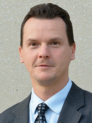
Presenters during New Tools to Visualize Autoimmune Tissue Targets, which will take place from 11:00 am – 12:00 pm in Room B309, Building B in the Georgia World Congress Center, include Janos Peti-Peterdi, MD, PhD, who will describe the interactions between immune cells and the local tissue environment in lupus nephritis using novel single cell imaging approaches. Dr. Peti-Peterdi, Professor of Physiology and Neuroscience and Medicine, and Director of the ZNI Multiphoton Core Facility at the University of Southern California Keck School of Medicine, also will review how cell imaging technologies are providing insights into lupus nephritis pathogenesis.
“Up to 60 percent of lupus patients will develop inflammation in the kidney, also known as lupus nephritis. When the kidneys are inflamed, they can’t function normally and can leak protein and if the inflammation is not controlled, it can lead to kidney failure,” Dr. Peti-Peterdi said. “Due to the unmet medical need for specific and highly efficient and mechanism-based new therapies, improved understanding of the disease pathomechanism is essential.”
Using a state-of-the-art intravital imaging technique developed by Dr. Peti-Peterdi’s research team, investigators are able to visualize and study the earliest stage of lupus development and understand the true sources of lupus complications.
“During this session, I will be presenting direct visual evidence from high-power intravital imaging of the local kidney tissue microenvironment in new animal models, showing that an interaction between circulating immune cells and an altered glomerular endothelium plays central roles in the development of lupus nephritis,” Dr. Peti-Peterdi said.
This finding may guide the future development of new targeted drugs and treatments for lupus nephritis. Based on targeting the molecular mechanisms that control a newly discovered tissue repair process, the Peti-Peterdi lab is currently developing new regenerative therapeutic approaches for the treatment of lupus nephritis.
“In our early pre-clinical work, we successfully validated a new therapeutic strategy to regenerate the damaged kidney filters and preserve renal function, and to address lupus complications in vital organs by targeting damaged cells and clearing clogged blood vessels,” Dr. Peti-Peterdi said.
Also during this session, Sean Bendall, PhD, Assistant Professor (Research) of Pathology at Stanford University, will review how novel high content imaging approaches, such as multiplexed ion beam imaging (MIBI), may be used to define cellular interactions in tissue at the single cell level in health and disease.
